| 中文名稱 | 叉頭蛋白P3抗體 |
| 別 名 | AIID; AIID; DIETER; DIETER; Forkhead box P3; Forkhead box protein P3; FOXP3_HUMAN; FOXP3delta7; Immune dysregulation polyendocrinopathy enteropathy X linked; Immunodeficiency polyendocrinopathy enteropathy X linked; IPEX; IPEX; JM2; JM2; MGC141961; MGC141963; OTTHUMP00000025832; OTTHUMP00000025833; OTTHUMP00000226737; PIDX; PIDX; SCURFIN; SCURFIN; XPID; XPID. |
| 研究領域 | 轉錄調節(jié)因子 |
| 抗體來源 | Rabbit |
| 克隆類型 | Polyclonal |
| 交叉反應 | Human, Mouse, Rat, (predicted: Dog, Pig, Cow, Horse, Rabbit, Sheep, Guinea Pig, ) |
| 產品應用 | WB=1:500-2000 ELISA=1:500-1000 IHC-P=1:100-500 IHC-F=1:100-500 Flow-Cyt=0.2ug/Test ICC=1:100-500 IF=1:100-500 (石蠟切片需做抗原修復) not yet tested in other applications. optimal dilutions/concentrations should be determined by the end user. |
| 分 子 量 | 47kDa |
| 細胞定位 | 細胞核 |
| 性 狀 | Liquid |
| 濃 度 | 1mg/ml |
| 免 疫 原 | KLH conjugated synthetic peptide derived from human FoxP3:331-431/431 |
| 亞 型 | IgG |
| 純化方法 | affinity purified by Protein A |
| 儲 存 液 | 0.01M TBS(pH7.4) with 1% BSA, 0.03% Proclin300 and 50% Glycerol. |
| 保存條件 | Shipped at 4℃. Store at -20 °C for one year. Avoid repeated freeze/thaw cycles. |
| PubMed | PubMed |
| 產品介紹 | The protein encoded by this gene is a member of the forkhead/winged-helix family of transcriptional regulators. Defects in this gene are the cause of immunodeficiency polyendocrinopathy, enteropathy, X-linked syndrome (IPEX), also known as X-linked autoimmunity-immunodeficiency syndrome. Alternatively spliced transcript variants encoding different isoforms have been identified. [provided by RefSeq, Jul 2008]. Function: Probable transcription factor. Plays a critical role in the control of immune response. Subunit: Interacts with IKZF3. Subcellular Location: Nucleus (Potential). Post-translational modifications: Acetylation on lysine residues stabilizes FOXP3 and promotes differentiation of T-cells into induced regulatory T-cells (iTregs) associated with suppressive functions. Deacetylated by SIRT1. DISEASE: Defects in FOXP3 are the cause of immunodeficiency polyendocrinopathy, enteropathy, X-linked syndrome (IPEX) [MIM:304790]; also known as X-linked autoimmunity-immunodeficiency syndrome. IPEX is characterized by neonatal onset insulin-dependent diabetes mellitus, infections, secretory diarrhea, trombocytopenia, anemia and eczema. It is usually lethal in infancy. Similarity: Contains 1 C2H2-type zinc finger. Contains 1 fork-head DNA-binding domain. SWISS: Q9BZS1 Gene ID: 50943 Database links: Entrez Gene: 50943 Human Entrez Gene: 20371 Mouse Entrez Gene: 317382 Rat Omim: 300292 Human SwissProt: Q9BZS1 Human SwissProt: Q99JB6 Mouse SwissProt: D3ZKI1 Rat Unigene: 247700 Human Unigene: 182291 Mouse Important Note: This product as supplied is intended for research use only, not for use in human, therapeutic or diagnostic applications. |
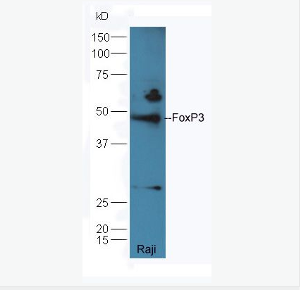
| 產品圖片 | 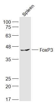 Sample: Sample:Spleen (Rat) Lysate at 40 ug Primary: Anti-FoxP3 (bs-10211R) at 1/300 dilution Secondary: IRDye800CW Goat Anti-Rabbit IgG at 1/20000 dilution Predicted band size: 47 kD Observed band size: 47 kD 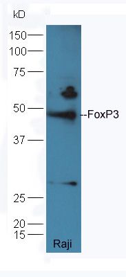 Protein: Raji(human) lysates at 40ug; Protein: Raji(human) lysates at 40ug;Primary: rabbit Anti-FoxP3 (bs-10211R) at 1:300; Secondary: HRP conjugated Goat-Anti-rabbit IgG(bs-0295G-HRP) at 1: 5000; Predicted band size:47 kD Observed band size:47 kD 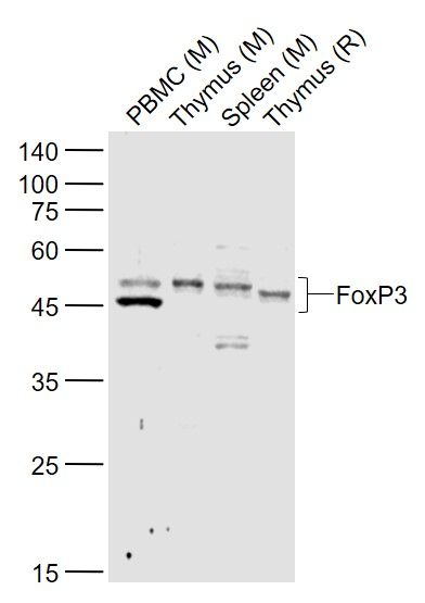 Sample: Sample:Lane 1: PBMC (Mouse) Lysate at 40 ug Lane 2: Thymus (Mouse) Lysate at 40 ug Lane 3: Spleen (Mouse) Lysate at 40 ug Lane 4: Thymus (Rat) Lysate at 40 ug Primary: Anti-FoxP3 (bs-10211R) at 1/1000 dilution Secondary: IRDye800CW Goat Anti-Rabbit IgG at 1/20000 dilution Predicted band size: 43/45 kD Observed band size: 43/45 kD 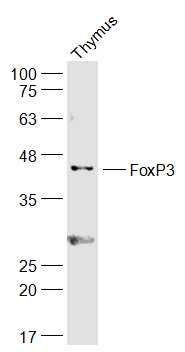 Sample: Sample:Thymus (Mouse) Lysate at 40 ug Primary: Anti-FoxP3 (10211R) at 1/300 dilution Secondary: IRDye800CW Goat Anti-Rabbit IgG at 1/20000 dilution Predicted band size: 47 kD Observed band size: 47 kD 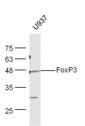 Sample: Sample:U937 Cell (Human) Lysate at 30 ug Primary: Anti-FoxP3 (Bs-10211R) at 1/300 dilution Secondary: IRDye800CW Goat Anti-Rabbit IgG at 1/20000 dilution Predicted band size: 47 kD Observed band size: 48 kD 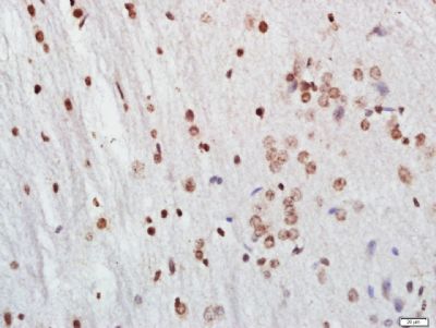 Tissue/cell: Rat brain tissue; 4% Paraformaldehyde-fixed and paraffin-embedded; Tissue/cell: Rat brain tissue; 4% Paraformaldehyde-fixed and paraffin-embedded;Antigen retrieval: citrate buffer ( 0.01M, pH 6.0 ), Boiling bathing for 15min; Block endogenous peroxidase by 3% Hydrogen peroxide for 30min; Blocking buffer (normal goat serum,C-0005) at 37℃ for 20 min; Incubation: Anti-FoxP3 Polyclonal Antibody, Unconjugated(10211R) 1:500, overnight at 4°C, followed by conjugation to the secondary antibody(SP-0023) and DAB(C-0010) staining 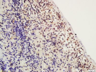 Tissue/cell: rat spleen; 4% Paraformaldehyde-fixed and paraffin-embedded; Tissue/cell: rat spleen; 4% Paraformaldehyde-fixed and paraffin-embedded;Antigen retrieval: citrate buffer ( 0.01M, pH 6.0 ), Boiling bathing for 15min; Block endogenous peroxidase by 3% Hydrogen peroxide for 30min; Blocking buffer (normal goat serum,C-0005) at 37℃ for 20 min; Incubation: Anti-FoxP3 Polyclonal Antibody, Unconjugated(bs-10211R) 1:200, overnight at 4°C, followed by conjugation to the secondary antibody(SP-0023) and DAB(C-0010) staining 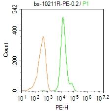 Blank control: Mouse spleen. Blank control: Mouse spleen.Primary Antibody (green line): Rabbit Anti-FoxP3/PE Conjugated antibody (10211R-PE) Dilution: 0.2μg /10^6 cells; Isotype Control Antibody (orange line): Rabbit IgG-PE . Protocol The cells were fixed with 4% PFA (10min at room temperature)and then permeabilized with 90% ice-cold methanol for 20 min at-20℃. The cells were then incubated in 5% BSA to block non-specific protein-protein interactions for 30 min at room temperature. The cells were stained with Primary Antibody for 30 min at room temperature. Acquisition of 20,000 events was performed. 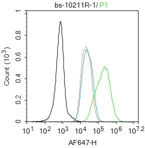 Blank control: MCF7. Blank control: MCF7.Primary Antibody (green line): Rabbit Anti-FoxP3 antibody (bs-10211R) Dilution: 2μg /10^6 cells; Isotype Control Antibody (orange line): Rabbit IgG . Secondary Antibody : Goat anti-rabbit IgG-AF647 Dilution: 1μg /test. Protocol The cells were fixed with 4% PFA (10min at room temperature)and then permeabilized with 90% ice-cold methanol for 20 min at-20℃.The cells were then incubated in 5%BSA to block non-specific protein-protein interactions for 30 min at room temperature .Cells stained with Primary Antibody for 30 min at room temperature. The secondary antibody used for 40 min at room temperature. Acquisition of 20,000 events was performed. 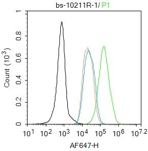 Blank control: MCF7. Blank control: MCF7.Primary Antibody (green line): Rabbit Anti-FoxP3 antibody (bs-10211R) Dilution: 2μg /10^6 cells; Isotype Control Antibody (orange line): Rabbit IgG . Secondary Antibody : Goat anti-rabbit IgG-AF647 Dilution: 1μg /test. Protocol The cells were fixed with 4% PFA (10min at room temperature)and then permeabilized with 90% ice-cold methanol for 20 min at-20℃.The cells were then incubated in 5%BSA to block non-specific protein-protein interactions for 30 min at room temperature .Cells stained with Primary Antibody for 30 min at room temperature. The secondary antibody used for 40 min at room temperature. Acquisition of 20,000 events was performed. 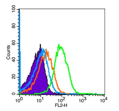 Blank control (Black line):Mouse spleen (Black). Blank control (Black line):Mouse spleen (Black).Primary Antibody (green line): Rabbit Anti-FoxP3 antibody (bs-10211R) Dilution: 3μg /10^6 cells; Isotype Control Antibody (orange line): Rabbit IgG . Secondary Antibody (white blue line): Goat anti-rabbit IgG-PE Dilution: 1μg /test. Protocol The cells were fixed with 4% PFA (10min at room temperature)and then permeabilized with 90% ice-cold methanol for 20 min at room temperature. The cells were then incubated in 5%BSA goat serum to block non-specific protein-protein interactions for 15 min at room temperature .Cells stained with Primary Antibody for 30 min at room temperature. The secondary antibody used for 40 min at room temperature. Acquisition of 20,000 events was performed. |
我要詢價
*聯(lián)系方式:
(可以是QQ、MSN、電子郵箱、電話等,您的聯(lián)系方式不會被公開)
*內容:


