| 中文名稱 | 叉頭蛋白N1抗體 |
| 別 名 | FKHL20; Forkhead box N1; Forkhead box protein N1; FOXN 1; FOXN1; FOXN1_HUMAN; RONU; Rowett nude; Transcription factor winged-helix nude; WHN; Winged helix nude; Winged-helix transcription factor nude. |
| 研究領(lǐng)域 | 細胞生物 免疫學 染色質(zhì)和核信號 轉(zhuǎn)錄調(diào)節(jié)因子 表觀遺傳學 |
| 抗體來源 | Rabbit |
| 克隆類型 | Polyclonal |
| 交叉反應(yīng) | Human, Mouse, Rat, (predicted: Chicken, Dog, Pig, Cow, Horse, Rabbit, ) |
| 產(chǎn)品應(yīng)用 | WB=1:500-2000 ELISA=1:500-1000 IHC-P=1:100-500 IHC-F=1:100-500 Flow-Cyt=1ug/Test IF=1:50-200 (石蠟切片需做抗原修復(fù)) not yet tested in other applications. optimal dilutions/concentrations should be determined by the end user. |
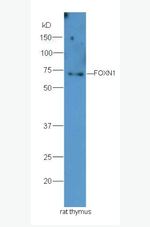
| 分 子 量 | 69kDa |
| 細胞定位 | 細胞核 |
| 性 狀 | Liquid |
| 濃 度 | 1mg/ml |
| 免 疫 原 | KLH conjugated synthetic peptide derived from human FOXN1:321-420/648 |
| 亞 型 | IgG |
| 純化方法 | affinity purified by Protein A |
| 儲 存 液 | 0.01M TBS(pH7.4) with 1% BSA, 0.03% Proclin300 and 50% Glycerol. |
| 保存條件 | Shipped at 4℃. Store at -20 °C for one year. Avoid repeated freeze/thaw cycles. |
| PubMed | PubMed |
| 產(chǎn)品介紹 | Mutations in the winged-helix transcription factor gene at the nude locus in mice and rats produce the pleiotropic phenotype of hairlessness and athymia, resulting in a severely compromised immune system. This gene is orthologous to the mouse and rat genes and encodes a similar DNA-binding transcription factor that is thought to regulate keratin gene expression. A mutation in this gene has been correlated with T-cell immunodeficiency, the skin disorder congenital alopecia, and nail dystrophy. Alternative splicing in the 5' UTR of this gene has been observed. [provided by RefSeq, Jul 2008]. Function: Transcriptional regulator involved in development. Subcellular Location: Nucleus. Tissue Specificity: Expressed in thymus. Similarity: Contains 1 fork-head DNA-binding domain. SWISS: O15353 Gene ID: 8456 Database links: Entrez Gene: 8456 Human Omim: 600838 Human SwissProt: O15353 Human Unigene: 663679 Human Important Note: This product as supplied is intended for research use only, not for use in human, therapeutic or diagnostic applications. |
| 產(chǎn)品圖片 | 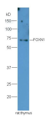 Sample: Thymus (Rat) Lysate at 40 ug Sample: Thymus (Rat) Lysate at 40 ugPrimary: Anti-FOXN1 (bs-6970R) at 1/300 dilution Secondary: HRP conjugated Goat-Anti-rabbit IgG (bs-0295G-HRP) at 1/5000 dilution Predicted band size: 69 kD Observed band size: 69 kD 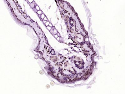 Paraformaldehyde-fixed, paraffin embedded (Mouse skin); Antigen retrieval by boiling in sodium citrate buffer (pH6.0) for 15min; Block endogenous peroxidase by 3% hydrogen peroxide for 20 minutes; Blocking buffer (normal goat serum) at 37°C for 30min; Antibody incubation with (FOXN1) Polyclonal Antibody, Unconjugated (bs-6970R) at 1:400 overnight at 4°C, followed by operating according to SP Kit(Rabbit) (sp-0023) instructionsand DAB staining. Paraformaldehyde-fixed, paraffin embedded (Mouse skin); Antigen retrieval by boiling in sodium citrate buffer (pH6.0) for 15min; Block endogenous peroxidase by 3% hydrogen peroxide for 20 minutes; Blocking buffer (normal goat serum) at 37°C for 30min; Antibody incubation with (FOXN1) Polyclonal Antibody, Unconjugated (bs-6970R) at 1:400 overnight at 4°C, followed by operating according to SP Kit(Rabbit) (sp-0023) instructionsand DAB staining.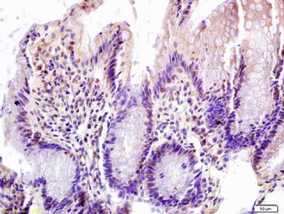 Tissue/cell: rat colon tissue; 4% Paraformaldehyde-fixed and paraffin-embedded; Tissue/cell: rat colon tissue; 4% Paraformaldehyde-fixed and paraffin-embedded;Antigen retrieval: citrate buffer ( 0.01M, pH 6.0 ), Boiling bathing for 15min; Block endogenous peroxidase by 3% Hydrogen peroxide for 30min; Blocking buffer (normal goat serum,C-0005) at 37℃ for 20 min; Incubation: Anti-FOXN1 Polyclonal Antibody, Unconjugated(bs-6970R) 1:200, overnight at 4°C, followed by conjugation to the secondary antibody(SP-0023) and DAB(C-0010) staining 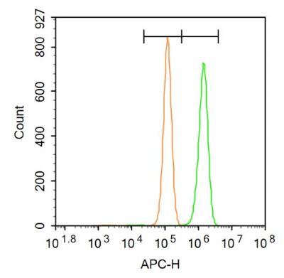 Blank control: Hela. Blank control: Hela.Primary Antibody (green line): Rabbit Anti-FOXN1 antibody (bs-6970R) Dilution: 1μg /10^6 cells; Isotype Control Antibody (orange line): Rabbit IgG . Secondary Antibody: Goat anti-rabbit IgG-AF647 Dilution: 1μg /test. Protocol The cells were fixed with 4% PFA (10min at room temperature)and then permeabilized with 90% ice-cold methanol for 20 min at room temperature. The cells were then incubated in 5%BSA to block non-specific protein-protein interactions for 30 min at room temperature .Cells stained with Primary Antibody for 30 min at room temperature. The secondary antibody used for 40 min at room temperature. Acquisition of 20,000 events was performed. 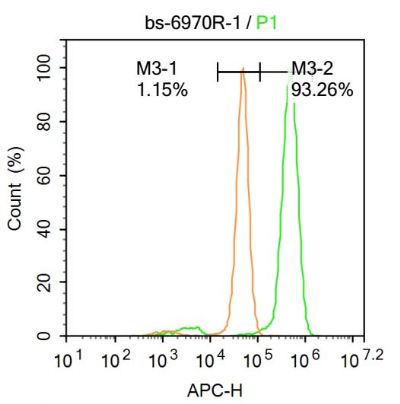 Blank control (Black line): Hela (Black). Blank control (Black line): Hela (Black).Primary Antibody (green line): Rabbit Anti-FOXN1 antibody (bs-6970R) Dilution: 1μg /10^6 cells; Isotype Control Antibody (orange line): Rabbit IgG . Secondary Antibody (white blue line): Goat anti-rabbit IgG-AF647 Dilution: 1μg /test. Protocol The cells were fixed with 4% PFA (10min at room temperature)and then permeabilized with 90% ice-cold methanol for 20 min at room temperature. The cells were then incubated in 5%BSA to block non-specific protein-protein interactions for 30 min at -20℃ .Cells stained with Primary Antibody for 30 min at room temperature. The secondary antibody used for 40 min at room temperature. Acquisition of 20,000 events was performed. |
我要詢價
*聯(lián)系方式:
(可以是QQ、MSN、電子郵箱、電話等,您的聯(lián)系方式不會被公開)
*內(nèi)容:


