Entrez Gene: 3654 Human Entrez Gene: 16179 Mouse Entrez Gene: 363520 Rat Omim: 300283 Human SwissProt: P51617 Human SwissProt: Q62406 Mouse Unigene: 522819 Human Unigene: 38241 Mouse Unigene: 22238 Rat
中文名稱 磷酸化白介素-1受體相關(guān)激酶1抗體 別 名 IRAK (phospho T209); p-IRAK (phospho T209); IRAK1 (Phospho-Thr209); IRAK1 (Phospho-T209); IRAK1 (p-Thr209); Il1rak; Il1rak; Interleukin 1 receptor associated kinase 1; Interleukin 1 receptor associated kinase 1; Interleukin 1 receptor associated kinase 2; Interleukin-1 receptor-associated kinase 1; IRAK; IRAK-1; IRAK1; IRAK1; IRAK1_HUMAN; IRAK2; IRAK2; mPLK; mPLK; Pelle; Pelle; Pelle homolog; Pelle-like protein kinase; Plpk. 產(chǎn)品類型 磷酸化抗體 研究領(lǐng)域 腫瘤 免疫學(xué) 細(xì)胞凋亡 轉(zhuǎn)錄調(diào)節(jié)因子 抗體來(lái)源 Rabbit 克隆類型 Polyclonal 交叉反應(yīng) Human, Rat, 產(chǎn)品應(yīng)用 WB=1:500-2000 IHC-P=1:100-500 Flow-Cyt=0.2μg /Test (石蠟切片需做抗原修復(fù))
not yet tested in other applications.
optimal dilutions/concentrations should be determined by the end user. 分 子 量 78kDa 細(xì)胞定位 細(xì)胞核 細(xì)胞漿 性 狀 Liquid 濃 度 1mg/ml 免 疫 原 KLH conjugated synthesised phosphopeptide derived from human IRAK1 around the phosphorylation site of Thr209:RG(p-T)HN 亞 型 IgG 純化方法 affinity purified by Protein A 儲(chǔ) 存 液 0.01M TBS(pH7.4) with 1% BSA, 0.03% Proclin300 and 50% Glycerol. 保存條件 Shipped at 4℃. Store at -20 °C for one year. Avoid repeated freeze/thaw cycles. PubMed PubMed 產(chǎn)品介紹 This gene encodes the interleukin-1 receptor-associated kinase 1, one of two putative serine/threonine kinases that become associated with the interleukin-1 receptor (IL1R) upon stimulation. This gene is partially responsible for IL1-induced upregulation of the transcription factor NF-kappa B. Alternatively spliced transcript variants encoding different isoforms have been found for this gene. [provided by RefSeq, Jul 2008]
Function:
Serine/threonine-protein kinase that plays a critical role in initiating innate immune response against foreign pathogens. Involved in Toll-like receptor (TLR) and IL-1R signaling pathways. Is rapidly recruited by MYD88 to the receptor-signaling complex upon TLR activation. Association with MYD88 leads to IRAK1 phosphorylation by IRAK4 and subsequent autophosphorylation and kinase activation. Phosphorylates E3 ubiquitin ligases Pellino proteins (PELI1, PELI2 and PELI3) to promote pellino-mediated polyubiquitination of IRAK1. Then, the ubiquitin-binding domain of IKBKG/NEMO binds to polyubiquitinated IRAK1 bringing together the IRAK1-MAP3K7/TAK1-TRAF6 complex and the NEMO-IKKA-IKKB complex. In turn, MAP3K7/TAK1 activates IKKs (CHUK/IKKA and IKBKB/IKKB) leading to NF-kappa-B nuclear translocation and activation. Alternatively, phosphorylates TIRAP to promote its ubiquitination and subsequent degradation. Phosphorylates the interferon regulatory factor 7 (IRF7) to induce its activation and translocation to the nucleus, resulting in transcriptional activation of type I IFN genes, which drive the cell in an antiviral state. When sumoylated, translocates to the nucleus and phosphorylates STAT3
Subunit:
Homodimer. Interacts with TOLLIP; this interaction occurs in the cytosol prior to receptor activation. Interacts with MYD88; this interaction recruits IRAK1 to the stimulated receptor complex. Interacts with IL1RL1. Interacts with IRAK1BP1. Associates with TRAF6, PELI1 and IRAK4; this complex recruits MAP3K7/TAK1, TAB1 and TAB2 to mediate NF-kappa-B activation. Interacts (when polyubiquitinated) with IKBKG/NEMO.
Subcellular Location:
Cytoplasm. Nucleus. Note=Translocates to the nucleus when sumoylated.
Tissue Specificity:
Isoform 1 and isoform 2 are ubiquitously expressed in all tissues examined, with isoform 1 being more strongly expressed than isoform 2.
Post-translational modifications:
Following recruitment on the activated receptor complex, phosphorylated on Thr-209, probably by IRAK4, resulting in a conformational change of the kinase domain, allowing further phosphorylations to take place. Thr-387 phosphorylation in the activation loop is required to achieve full enzymatic activity.
Polyubiquitinated after cell stimulation with IL-1-beta by PELI1, PELI2 and PELI3. Polyubiquitination occurs with polyubiquitin chains linked through 'Lys-63'. Ubiquitination promotes interaction with NEMO/IKBKG. Also sumoylated; leading to nuclear translocation.
Similarity:
Belongs to the protein kinase superfamily. TKL Ser/Thr protein kinase family. Pelle subfamily.
Contains 1 death domain.
Contains 1 protein kinase domain.
SWISS:
P51617
Gene ID:
3654
Database links:
Important Note:
This product as supplied is intended for research use only, not for use in human, therapeutic or diagnostic applications.
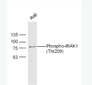
產(chǎn)品圖片 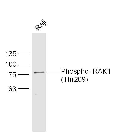 Sample: Raji Cell Lysate at 30 ug
Primary: Anti- phospho-IRAK1(Thr209) (bs-3193R) at 1/300 dilution
Secondary: IRDye800CW Goat Anti-Rabbit IgG at 1/20000 dilution
Predicted band size: 78 kD
Observed band size: 78 kD
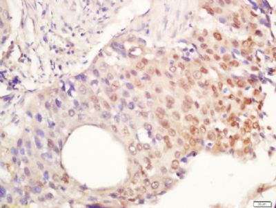 Tissue/cell: Human lung cancer tissue; 4% Paraformaldehyde-fixed and paraffin-embedded;
Antigen retrieval: citrate buffer ( 0.01M, pH 6.0 ), Boiling bathing for 15min; Block endogenous peroxidase by 3% Hydrogen peroxide for 30min; Blocking buffer (normal goat serum,C-0005) at 37℃ for 20 min;
Incubation: Anti-Phospho-IRAK1 (Ser376) Polyclonal Antibody, Unconjugated(bs-3193R) 1:200, overnight at 4°C, followed by conjugation to the secondary antibody(SP-0023) and DAB(C-0010) staining
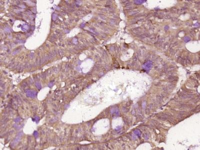 Paraformaldehyde-fixed, paraffin embedded (human cervical carcinoma); Antigen retrieval by boiling in sodium citrate buffer (pH6.0) for 15min; Block endogenous peroxidase by 3% hydrogen peroxide for 20 minutes; Blocking buffer (normal goat serum) at 37°C for 30min; Antibody incubation with (IRAK1 (Thr209)) Polyclonal Antibody, Unconjugated (bs-3193R) at 1:400 overnight at 4°C, followed by operating according to SP Kit(Rabbit) (sp-0023) instructionsand DAB staining.
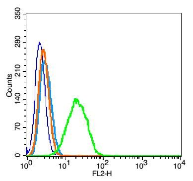 Blank control(blue): RSC96 cells (fixed with 2% paraformaldehyde (10 min) , then permeabilized with 90% ice-cold methanol for 30 min on ice).
Primary Antibody:Rabbit Anti-Phospho-IRAK1 (Thr209) antibody(bs-3193R), Dilution: 0.2μg in 100 μL 1X PBS containing 0.5% BSA;
Isotype Control Antibody: Rabbit IgG(orange),used under the same conditions );
Secondary Antibody: Goat anti-rabbit IgG-PE(white blue), Dilution: 1:200 in 1 X PBS containing 0.5% BSA.
您好,歡迎光臨上海雅吉生物商城!


