

| 中文名稱 | CD3/CD3-ε 抗體 |
| 別 名 | CD3 epsilon; CD3e; CD3e antigen epsilon polypeptide (TiT3 complex); CD3E antigen epsilon polypeptide; CD3E antigen, epsilon subunit; CD3e molecule epsilon; CD3e molecule, epsilon (CD3 TCR complex); CD3e molecule, epsilon (CD3-TCR complex); CD3E_HUMAN; IMD18; T cell antigen receptor complex epsilon subunit of T3; T cell surface antigen T3/Leu 4 epsilon chain; T cell surface glycoprotein CD3 epsilon chain; T-cell surface antigen T3/Leu-4 epsilon chain; T-cell surface glycoprotein CD3 epsilon chain; T3E; TCRE. |
| 研究領(lǐng)域 | 腫瘤 細(xì)胞生物 免疫學(xué) 干細(xì)胞 細(xì)胞膜受體 細(xì)胞表面分子 淋巴細(xì)胞 t-淋巴細(xì)胞 |
| 抗體來(lái)源 | Rabbit |
| 克隆類型 | Polyclonal |
| 交叉反應(yīng) | Human, Mouse, (predicted: Rat, MACFA) |
| 產(chǎn)品應(yīng)用 | ELISA=1:500-1000 IHC-P=1:100-500 IHC-F=1:100-500 Flow-Cyt=2μg/Test IF=1:100-500 (石蠟切片需做抗原修復(fù)) not yet tested in other applications. optimal dilutions/concentrations should be determined by the end user. |
| 分 子 量 | 20kDa |
| 細(xì)胞定位 | 細(xì)胞膜 |
| 性 狀 | Liquid |
| 濃 度 | 1mg/ml |
| 免 疫 原 | KLH conjugated synthetic peptide derived from mouse CD3E:101-189/189 |
| 亞 型 | IgG |
| 純化方法 | affinity purified by Protein A |
| 儲(chǔ) 存 液 | 0.01M TBS(pH7.4) with 1% BSA, 0.03% Proclin300 and 50% Glycerol. |
| 保存條件 | Shipped at 4℃. Store at -20 °C for one year. Avoid repeated freeze/thaw cycles. |
| PubMed | PubMed |
| 產(chǎn)品介紹 | The protein encoded by this gene is the CD3-epsilon polypeptide, which together with CD3-gamma, -delta and -zeta, and the T-cell receptor alpha/beta and gamma/delta heterodimers, forms the T-cell receptor-CD3 complex. This complex plays an important role in coupling antigen recognition to several intracellular signal-transduction pathways. The genes encoding the epsilon, gamma and delta polypeptides are located in the same cluster on chromosome 11. The epsilon polypeptide plays an essential role in T-cell development. Defects in this gene cause immunodeficiency. This gene has also been linked to a susceptibility to type I diabetes in women. [provided by RefSeq, Jul 2008]. Function: The CD3 complex mediates signal transduction. Subunit: The TCR/CD3 complex of T-lymphocytes consists of either a TCR alpha/beta or TCR gamma/delta heterodimer coexpressed at the cell surface with the invariant subunits of CD3 labeled gamma, delta, epsilon, zeta, and eta. Subcellular Location: Membrane; Single-pass type I membrane protein. Similarity: Contains 1 Ig-like (immunoglobulin-like) domain. Contains 1 ITAM domain. SWISS: P22646 Gene ID: 12501 Database links: Entrez Gene: 916 Human Entrez Gene: 12501 Mouse Omim: 186830 Human SwissProt: P07766 Human SwissProt: P22646 Mouse Unigene: 3003 Human Unigene: 210361 Mouse Important Note: This product as supplied is intended for research use only, not for use in human, therapeutic or diagnostic applications. |
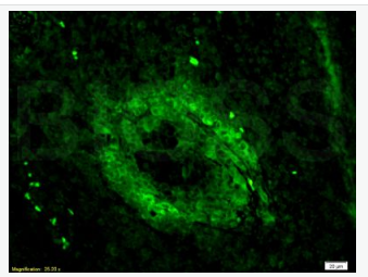
| 產(chǎn)品圖片 | 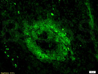 Tissue/cell: mouse lymphoma tissue;4% Paraformaldehyde-fixed and paraffin-embedded; Tissue/cell: mouse lymphoma tissue;4% Paraformaldehyde-fixed and paraffin-embedded;Antigen retrieval: citrate buffer ( 0.01M, pH 6.0 ), Boiling bathing for 15min; Blocking buffer (normal goat serum,C-0005) at 37℃ for 20 min; Incubation: Anti-CD3 Polyclonal Antibody, Unconjugated(bs-0765R) 1:200, overnight at 4°C; The secondary antibody was Goat Anti-Rabbit IgG, FITC conjugated(bs-0295G-FITC)used at 1:200 dilution for 40 minutes at 37°C. 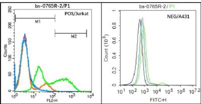 Black line : Positive blank control (Jurkat); Negative blank control (A431) Black line : Positive blank control (Jurkat); Negative blank control (A431)Green line : Primary Antibody (Rabbit Anti- CD3E antibody (bs-0765R) ) Orange line:Isotype Control Antibody (Rabbit IgG) . Blue line : Secondary Antibody (Goat anti-rabbit IgG-PE)/(Goat anti-rabbit IgG-PE) Jurkat(Positive)and A431(Negative control)cells (black) were incubated in 5% BSA blocking buffer for 30 min at room temperature. Cells were then stained with CD3E Antibody(bs-0765R)at 1:50 dilution in blocking buffer and incubated for 30 min at room temperature, washed twice with 2% BSA in PBS, followed by secondary antibody(blue) incubation for 40 min at room temperature. Acquisitions of 20,000 events were performed. Cells stained with primary antibody (green), and isotype control (orange). 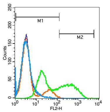 Blank control: Jurkat cells(blue). Blank control: Jurkat cells(blue).Primary Antibody:Rabbit Anti-CD3E antibody(bs-0765R), Dilution: 3μg in 100 μL 1X PBS containing 0.5% BSA; Isotype Control Antibody: Rabbit IgG(orange) ,used under the same conditions ); Secondary Antibody: Goat anti-rabbit IgG-PE(white blue), Dilution: 1:200 in 1 X PBS containing 0.5% BSA. Protocol The cells were fixed with 2% paraformaldehyde (10 min) . Primary antibody (bs-0765R, 3μg /1x10^6 cells) were incubated for 30 min on the ice, followed by 1 X PBS containing 0.5% BSA + 1 0% goat serum (15 min) to block non-specific protein-protein interactions. Then the Goat Anti-rabbit IgG/PE antibody was added into the blocking buffer mentioned above to react with the primary antibody at 1/200 dilution for 30 min on ice. Acquisition of 20,000 events was performed. |
我要詢價(jià)
*聯(lián)系方式:
(可以是QQ、MSN、電子郵箱、電話等,您的聯(lián)系方式不會(huì)被公開(kāi))
*內(nèi)容: