| 產(chǎn)品編號 | YS-1615R |
| 英文名稱 | Cathepsin D |
| 中文名稱 | 組織蛋白酶D輕鏈抗體 |
| 研究領域 | 細胞生物 免疫學 神經(jīng)生物學 合成與降解 細胞粘附分子 細胞外基質(zhì) |
| 抗體來源 | Rabbit |
| 克隆類型 | Polyclonal |
| 交叉反應 | Rat, (predicted: Human, Mouse, Dog, Pig, Cow, Rabbit, ) |
| 產(chǎn)品應用 | WB=1:500-2000 ELISA=1:5000-10000 IHC-P=1:100-500 IHC-F=1:100-500 IF=1:100-500 (石蠟切片需做抗原修復) not yet tested in other applications. optimal dilutions/concentrations should be determined by the end user. |
| 理論分子量 | 11/38/45kDa |
| 細胞定位 | 細胞漿 分泌型蛋白 |
| 性 狀 | Liquid |
| 濃 度 | 1mg/ml |
| 免 疫 原 | KLH conjugated synthetic peptide derived from human Cathepsin D light chain: 101-200/412 |
| 亞 型 | IgG |
| 純化方法 | affinity purified by Protein A |
| 緩 沖 液 | 0.01M TBS(pH7.4) with 1% BSA, 0.03% Proclin300 and 50% Glycerol. |
| 保存條件 | Shipped at 4℃. Store at -20 °C for one year. Avoid repeated freeze/thaw cycles. |
| 注意事項 | This product as supplied is intended for research use only, not for use in human, therapeutic or diagnostic applications. |
| PubMed | PubMed |
| 產(chǎn)品介紹 | Cathepsin D is a normal lysosomal protease that is expressed in all cells. It is an aspartyl protease with a pH optimum in the range of 3-5, and contains two N-linked oligosaccharides. Cathepsin D is synthesized as an inactive 52 kDa pro enzyme. Activation involves the proteolytic removal of the 43 amino acid profragment and an internal cleavage to generate the two-chain form made up of 34 and 14 kDa subunits. Cathepsin D contains the mannose-6-phosphate lysosomal localization signal that targets the enzyme to the lysosomal compartment where it functions in the normal degradation of proteins. In certain tumor cells, Cathepsin D is abnormally processed and is secreted in its 52 kDa precursor form. Numerous clinical studies as well as in vitro evidence suggest that cathepsin D plays an important role in malignant transformation and may be a useful prognostic indicator for breast cancer and possibly Alzheimer's disease. Function: Acid protease active in intracellular protein breakdown. Involved in the pathogenesis of several diseases such as breast cancer and possibly Alzheimer disease. Subcellular Location: Lysosome. Melanosome. Identified by mass spectrometry in melanosome fractions from stage I to stage IV. Tissue Specificity: Expressed in the aorta extrcellular space (at protein level). Post-translational modifications: N- and O-glycosylated. DISEASE: Defects in CTSD are the cause of neuronal ceroid lipofuscinosis type 10 (CLN10); also known as neuronal ceroid lipofuscinosis due to cathepsin D deficiency. A form of neuronal ceroid lipofuscinosis with onset at birth or early childhood. Neuronal ceroid lipofuscinoses are progressive neurodegenerative, lysosomal storage diseases characterized by intracellular accumulation of autofluorescent liposomal material, and clinically by seizures, dementia, visual loss, and/or cerebral atrophy. Similarity: Belongs to the peptidase A1 family. SWISS: P07339 Gene ID: 1509 |
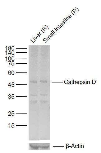
| 產(chǎn)品圖片 |  Sample: Lane 1: Rat Liver tissue lysates Lane 2: Rat Small intestine tissue lysates Primary: Anti-Cathepsin D (Ys-1615R) at 1/1000 dilution Secondary: IRDye800CW Goat Anti-Rabbit IgG at 1/20000 dilution Predicted band size: 11/38/45 kDa Observed band size: 46 kDa |
我要詢價
*聯(lián)系方式:
(可以是QQ、MSN、電子郵箱、電話等,您的聯(lián)系方式不會被公開)
*內(nèi)容:


